Category:Human pituitary gland
Jump to navigation
Jump to search
Subcategories
This category has the following 11 subcategories, out of 11 total.
D
H
- Hypophysial fossa (9 F)
- Hypothalamic–pituitary axis (2 F)
I
- Infundibular recess (15 F)
S
T
- Thyroid-stimulating hormone (3 F)
Media in category "Human pituitary gland"
The following 94 files are in this category, out of 94 total.
-
 01S-3A 022 ActivBil.ContraceptionHormonale.SchFoncMuet2.svg 1,286 × 909; 80 KB
-
 201405 pituitary gland in brain.png 400 × 400; 31 KB
-
 201405 pituitary gland.png 400 × 400; 36 KB
-
A-GRFa2Prop1Stem-(GPS)-Cell-Niche-in-the-Pituitary-pone.0004815.s005.ogv 15 s, 768 × 576; 1.72 MB
-
A-GRFa2Prop1Stem-(GPS)-Cell-Niche-in-the-Pituitary-pone.0004815.s006.ogv 15 s, 768 × 576; 3.35 MB
-
A-GRFa2Prop1Stem-(GPS)-Cell-Niche-in-the-Pituitary-pone.0004815.s007.ogv 17 s, 768 × 576; 1.09 MB
-
A-GRFa2Prop1Stem-(GPS)-Cell-Niche-in-the-Pituitary-pone.0004815.s008.ogv 1 min 22 s, 768 × 576; 4.09 MB
-
A-GRFa2Prop1Stem-(GPS)-Cell-Niche-in-the-Pituitary-pone.0004815.s009.ogv 1 min 5 s, 768 × 576; 2.91 MB
-
A-GRFa2Prop1Stem-(GPS)-Cell-Niche-in-the-Pituitary-pone.0004815.s010.ogv 33 s, 768 × 576; 2.39 MB
-
A-GRFa2Prop1Stem-(GPS)-Cell-Niche-in-the-Pituitary-pone.0004815.s011.ogv 20 s, 700 × 524; 2.56 MB
-

-
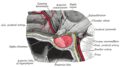
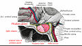
 Anatomy of the cavernous sinus.jpg 800 × 499; 93 KB
-
 Anterior and posterior pituitary.jpg 630 × 240; 28 KB
-
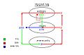
 Aufgabe der Hypophyse.svg 512 × 542; 187 KB
-
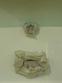 Basel 2012-10-05 Batch 2 (26).JPG 2,736 × 3,648; 3.03 MB
-
 Basel 2012-10-05 Batch 2 (27).JPG 2,736 × 3,648; 2.58 MB
-
 Basel 2012-10-05 Batch 2 (28).JPG 3,648 × 2,736; 3.68 MB
-
 Biosintesi della melatonina.JPG 378 × 960; 32 KB
-
 Corticotroph traces.png 2,481 × 1,057; 157 KB
-
De-Hypophyse.ogg 2.2 s; 21 KB
-
Development of the pituitary gland.webm 1 min 26 s, 640 × 480; 1.42 MB
-
Emergent-Synchronous-Bursting-of-Oxytocin-Neuronal-Network-pcbi.1000123.s001.ogv 20 s, 560 × 420; 1.85 MB
-
 Endocrine growth regulation.png 1,698 × 2,080; 1.05 MB
-
 Figure 28 01 07.JPG 1,058 × 713; 250 KB
-
 Gray1180-ar.png 672 × 375; 165 KB
-
 Gray1180.png 672 × 375; 45 KB
-
 Gray1181-ar.png 575 × 350; 127 KB
-
 Gray1181.png 575 × 350; 34 KB
-
 Gray516.png 600 × 681; 113 KB
-
 Gray721.png 600 × 333; 59 KB
-
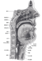 Gray994 zh.png 600 × 861; 329 KB
-
 Gray994.png 600 × 861; 112 KB
-
 Grays pituitary.png 600 × 333; 65 KB
-
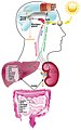
 Hipofisis-hormonas.jpg 896 × 814; 77 KB
-
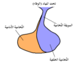
 Hipófise - Posterior.png 555 × 401; 174 KB
-
 Hipófise ou Glândula Pituitária.jpg 289 × 174; 7 KB
-
 Hipófisis-hormonas2.jpg 896 × 814; 80 KB
-
 Hormonas-hipofisis.jpg 896 × 814; 77 KB
-
 Hormone in der Pubertät.svg 512 × 390; 217 KB
-
 Human brain dura mater (reflections) description.JPG 700 × 486; 49 KB
-
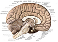
 Human brain left midsagitttal view closeup description 2.JPG 701 × 490; 61 KB
-
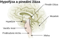 Hypofýza a pineální žláza.svg 114 × 76; 59 KB
-
 Hypophyse 2M - MR - 001.jpg 1,467 × 1,881; 292 KB
-
 Hypophyse 31jw postpartum - MRT T1 sag - 001.jpg 1,535 × 1,318; 123 KB
-
 Hypophyse et glandes pinéales.gif 400 × 259; 19 KB
-
 Hypophyse MRT sag.png 1,198 × 1,180; 615 KB
-
 Hypophyse und Epiphyse.jpg 656 × 512; 74 KB
-
 Hypophyse.png 2,252 × 2,072; 692 KB
-
 Hypophyseal gland.jpg 960 × 720; 87 KB
-
 Hypophysis.jpg 467 × 304; 16 KB
-
 Hypophysis3.gif 575 × 350; 39 KB
-
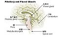 Illu pituitary pineal glands ja.JPG 400 × 255; 23 KB
-
 Illu pituitary pineal glands zh.jpg 400 × 259; 31 KB
-
 Illu pituitary pineal glands-az.png 400 × 259; 84 KB
-
 Illu pituitary pineal glands.jpg 400 × 259; 23 KB
-
 Innvervazione dell'epifisi.JPG 567 × 567; 27 KB
-

-

-
 Interplay between the central and peripheral circadian clocks.jpg 766 × 1,221; 127 KB
-
 L03 infundibulum 001.jpg 378 × 377; 30 KB
-
 Location of hypothalamus, pituitary gland and olfactory bulb. .gif 440 × 243; 14 KB
-
 LocationOfHypothalamus.jpg 350 × 250; 21 KB
-
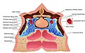
 Nasennebenhöhlen.gif 600 × 428; 66 KB
-
 Neurohipófisis.jpg 832 × 1,151; 166 KB
-
 Pineal Gland and Pituitary Body.jpg 291 × 170; 10 KB
-
 Pituitary development animation.gif 300 × 200; 52 KB
-
 Pituitary gland et vessel.jpg 700 × 600; 57 KB
-
 Pituitary gland image.png 800 × 455; 308 KB
-
 Pituitary gland representation.PNG 261 × 236; 8 KB
-
 Pituitary gland small.gif 200 × 200; 592 KB
-
 Pituitary gland-optic chiasm-sella turcica-es.png 625 × 347; 69 KB
-
 Pituitary gland-optic chiasm-sella turcica.jpg 600 × 333; 127 KB
-
 Pituitary gland.png 468 × 332; 210 KB
-
 Pituitary pineal glands.jpg 355 × 239; 22 KB
-
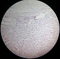 Pituitary slide.jpg 2,993 × 2,908; 3.51 MB
-
 Pituitary Stalk-ar.png 309 × 257; 9 KB
-
 Pituitary Stalk.png 309 × 257; 8 KB
-
 Pituitary Tumor Removal.png 1,500 × 750; 877 KB
-
 Pituitary xanthogranuloma.jpg 2,080 × 1,542; 857 KB
-
 Prolactinoma-art-zh.jpg 261 × 358; 50 KB
-
 Prolactinoma-art.jpg 261 × 358; 70 KB
-
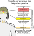 Regelmechanismus Körpertemperatur.svg 512 × 542; 240 KB
-
 Sajous's analytical cyclopædia of practical medicine (1904) (14778360432).jpg 1,554 × 2,282; 574 KB
-
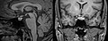
 Sella mri-contra.jpg 1,100 × 600; 112 KB
-
Sheehans Syndrome.webm 5 min 10 s, 1,920 × 1,080; 34.16 MB
-
 Silla turca TalloHipof.png 512 × 322; 309 KB
-
 Sistema porta H-H.jpg 1,226 × 716; 170 KB
-
 Sleep.PNG 537 × 455; 35 KB
-
 Slide2STE.JPG 960 × 720; 106 KB
-
 Sobo 1909 624 ar.png 3,060 × 2,247; 5.38 MB
-
 Sobo 1909 624.png 3,060 × 2,247; 19.71 MB
-
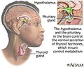 ThyroidImage.jpg 400 × 320; 68 KB
-
 Voluminoese Hypophyse in der Schwangerschaft 30W - MR T1 - 001.jpg 3,633 × 1,380; 280 KB
-
 Схема гипоталамо-эпифизарной системы.jpg 719 × 720; 145 KB


.JPG)
.JPG)
.JPG)


_description.JPG)


_(14765455831).jpg)


_(14778360432).jpg)


_(14768309272).jpg)